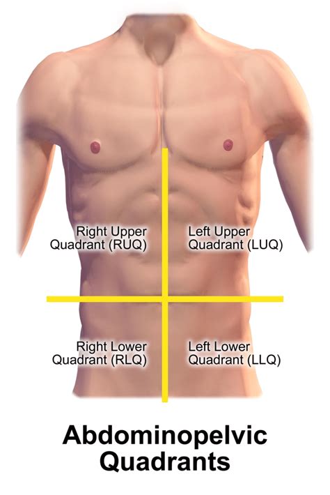
Understanding the quadrants of the abdomen is fundamental for medical professionals and even for laypersons to comprehend vital diagnostic insights. The abdomen is anatomically divided into four quadrants: the right upper quadrant (RUQ), left upper quadrant (LUQ), right lower quadrant (RLQ), and left lower quadrant (LLQ). This division helps in pinpointing the source of abdominal pain and assists in the assessment of various diseases and conditions. Let’s dive deeper into the specifics of each quadrant, offering actionable guidance to help both medical professionals and curious readers grasp these crucial concepts.
Why Understanding Abdominal Quadrants Matters
Mastering the anatomy and implications of each quadrant of the abdomen can be a lifesaver. For medical practitioners, identifying which quadrant is affected by symptoms like pain, swelling, or tenderness is crucial for effective diagnosis. For example, pain in the RUQ could point to issues with the liver or gallbladder, while pain in the LLQ might indicate appendicitis or problems with the large intestine. This guide aims to arm you with the knowledge to understand these critical regions and the potential conditions they encompass.
Here’s an overview of how this knowledge can be practically applied:
Quick Reference
- Immediate action item with clear benefit: Start with a thorough patient history and physical examination to determine the location of pain.
- Essential tip with step-by-step guidance: Perform a systematic examination of each quadrant using palpation to identify tenderness and note any masses or abnormal findings.
- Common mistake to avoid with solution: Assuming that right upper quadrant pain is always gallbladder-related without considering other possibilities, such as liver disease or pancreas issues.
Right Upper Quadrant (RUQ)
The RUQ includes a significant portion of the liver, the gallbladder, and part of the pancreas. Pain in this area is often indicative of liver conditions, gallbladder disease, or biliary tree issues. To make sense of RUQ pain, it’s vital to consider the following:
Actionable steps to diagnose RUQ symptoms:
- Begin with a thorough patient history, noting any previous abdominal issues or family history.
- Perform a physical examination, focusing on palpation to detect any liver enlargement or gallbladder tenderness.
- Order laboratory tests such as liver function tests (LFTs), complete blood count (CBC), and lipase levels to assess for possible liver or pancreatic conditions.
- Use imaging techniques like an abdominal ultrasound or CT scan to visualize the gallbladder and liver, helping to detect gallstones, liver enlargement, or other abnormalities.
Practical examples:
Consider a patient presenting with RUQ pain, nausea, and vomiting. The doctor might suspect biliary colic due to gallstones. An abdominal ultrasound would be the next step to confirm the presence of gallstones and to assess gallbladder wall thickening or pericholecystic fluid indicative of acute cholecystitis.
Left Upper Quadrant (LUQ)
The LUQ encompasses parts of the stomach, spleen, and left kidney, among other structures. Conditions affecting this quadrant might include splenic enlargement, kidney stones, or gastric issues.
Steps for diagnosing LUQ symptoms:
- Start with a detailed patient history, inquiring about any recent infections, trauma, or known renal problems.
- Conduct a focused physical examination, noting any signs of splenomegaly (enlarged spleen) or renal angle tenderness.
- Proceed with relevant lab tests such as a complete blood count (CBC) to check for splenomegaly and renal function tests to assess kidney health.
- Utilize imaging like an abdominal ultrasound or CT scan to better visualize splenic, renal, and gastric structures, detecting any anomalies.
Practical examples:
A patient might come in with LUQ pain, fatigue, and a palpable mass in the left upper abdomen. These symptoms may suggest splenomegaly due to conditions like lymphoma or mononucleosis. An ultrasound would help to confirm the spleen’s size and check for any abnormalities, guiding the need for further tests such as a bone marrow biopsy.
Right Lower Quadrant (RLQ)
The RLQ contains part of the small intestine, cecum, appendix, and the reproductive organs in both males and females. Conditions here might include appendicitis, ovarian cysts, or ectopic pregnancies in women.
Actionable steps to diagnose RLQ pain:
- Begin with a detailed history, focusing on the onset, duration, and character of pain, alongside associated symptoms like nausea, vomiting, and changes in bowel habits.
- Perform a physical examination, specifically looking for signs of appendicitis such as McBurney’s point tenderness and a positive Rovsing’s sign.
- Order laboratory tests including a complete blood count (CBC), which can show elevated white blood cells in appendicitis, and urine tests to rule out a urinary tract infection.
- Use imaging techniques such as an abdominal ultrasound, CT scan, or pelvic ultrasound to visualize the appendix and detect any signs of appendicitis or ovarian pathology.
Practical examples:
A patient presenting with RLQ pain, fever, and nausea could be diagnosed with appendicitis. An abdominal CT scan would be highly diagnostic, revealing a thickened appendix with surrounding inflammation. Surgical consultation would be warranted to prevent rupture and subsequent peritonitis.
Left Lower Quadrant (LLQ)
The LLQ houses the sigmoid colon, part of the small intestine, and the reproductive organs in females. It’s crucial to consider conditions like diverticulitis, ovarian cysts, or even constipation when evaluating LLQ pain.
Steps for diagnosing LLQ symptoms:
- Take a comprehensive history, asking about bowel habits, recent dietary changes, and any history of diverticulosis.
- Conduct a physical examination, noting any tenderness over the sigmoid colon and checking for signs of peritonitis.
- Order relevant lab tests such as a complete blood count (CBC) to check for signs of infection or inflammation and a stool test to check for gastrointestinal bleeding.
- Use imaging such as an abdominal ultrasound, CT scan, or contrast enema to visualize the colon and detect abnormalities like diverticula or masses.
Practical examples:
A patient experiencing LLQ pain, fever, and changes in bowel movements might be diagnosed with diverticulitis. A CT scan would help to confirm the diagnosis by showing inflamed diverticula and possibly abscess formation. Management would include antibiotics and, in severe cases, surgical intervention.
Practical FAQ
What are the common symptoms indicating a problem in each abdominal quadrant?
Symptoms vary depending on the quadrant involved:
- RUQ: Right upper quadrant pain may be associated with liver, gallbladder, or biliary tree issues such as right shoulder pain, nausea, vomiting, and jaundice.
- LUQ: Left upper quadrant pain may indicate spleen issues (splenomegaly), gastric problems, or kidney-related conditions, presenting with left shoulder pain and changes in blood count.
- RLQ: Right lower quadrant pain is often a hallmark of appendicitis, though it can also signal gynecological issues in females. Symptoms include fever, nausea, and pain radiating to the right lower abdomen.
- LLQ: Left lower quadrant pain could be due to diverticulitis, ovarian cysts in females, or constipation. Patients often report left-sided abdominal pain and changes in bowel habits.
Best Practices for Using Abdominal Quadrant Knowledge
Here are some best practices to follow when applying this knowledge in clinical settings:
- Always start with a thorough history and physical examination before any diagnostic tests. Understanding the patient’s full clinical picture is crucial.
- Utilize imaging techniques judiciously. While CT scans provide detailed images, they should be used when absolutely necessary due to radiation exposure.
- Consider the patient’s overall health and any comorbidities that could affect their presentation. For instance, a patient with diabetes might present with atypical symptoms.
- Comm